OCT technique and its applications
This technique is a novel and advanced method for imaging the surface and deeper layers of the eyes. With OCT, one can visualize the anatomic situation and even histologic status of the eyes. Indeed, unlike pathology which requires biopsy and staining tissues outside the body, OCT represents a slice of the tissue while it is live and in the human body with a high accuracy and quality. Since the major tissues of the eyes are transparent, this method of imaging has numerous applications in ophthalmology.
In OCT, a special laser light is irradiated to the eyes. Then, this light is reflected eventually after passing through different layers of the eyes. Then, the center of the device examines the way different layers of the eyes are organized by analyzing the light reflection, whereby it differentiates between normal and abnormal layers, and indicates the type of lesion with micron resolution and accuracy.
This type of imaging is mostly used for investigating the layers of retina and the optic nerve. However, there are also some types of OCT which can visualize the anterior layers of the eye including cornea, sclera, Iris, and lens. The difference between OCT and topography and conventional tomography is that topography is used for imaging the anterior layers of the eye including cornea. However, OCT is able to also investigate the posterior layers of the eye including retina. In addition, their optical and technology is also fundamentally different.
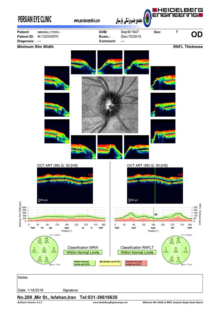
Application of OCT in glaucoma
Uses of OCT mostly include investigating the lesions and diseases of retina. In addition, OCT is also widely employed to diagnose and examine the course of glaucoma. Since in glaucoma, in response to increased ocular pressure, the neuronal layer of retina as well as the optic nerve become damaged, OCT pinpoints existence of damage and its magnitude, and as such it is considered a valuable instrument for early diagnosis examining the severity and course of the disease.
OCT angiography
Angiography is a type of imaging from blood vessels which precisely images the vessels after injecting a contrast agent. Recently, OCT has found application for this purpose. In OCT angiography, without any need to inject contrast agent or medications, the blood vessels of the retina and choroid are visualized carefully.
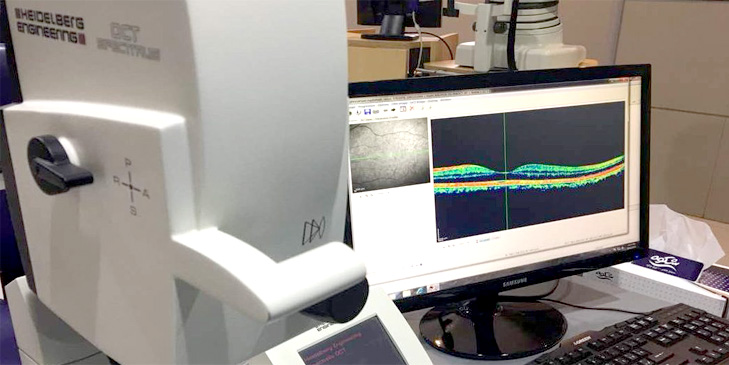
Parsian clinic was the first center in the province and among the first centers in the country and even the world which used this advanced technology.
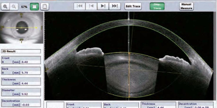
Anterior OCT (AS-OCT)
As stated earlier, OCT was first used to investigate the diseases of retina and glaucoma. However, today advanced OCT devices are used for investigating the cornea as well as the anterior chamber and angle of the eye. Using this technology, one can generate interesting and precise images of the structure of this part of the eye and measure the properties and dimensions of each part.
Parsian subspecialist ophthalmology clinic is proud to state that it has imported this technology into the province for the first time in 2006. Since then, this clinic has used advanced systems in this regard and currently it has the most advanced and complete set of posterior, anterior, and angiography OCT.
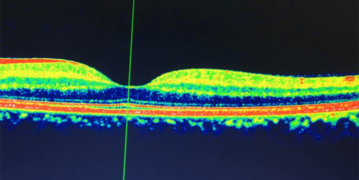
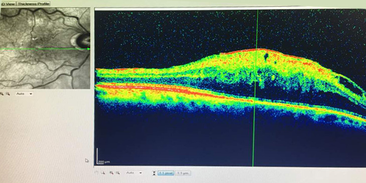
In the following figures, you observe a sample of normal retina, damaged retina, and the structure of the anterior part of the eyes as captured by OCT.