Topography and Tomography
are a kind of imaging used for
investigating the surface and depth of cornea, which represent all properties of the cornea including shape, unevenness, thickness, and corneal curvature. It also manifests the smoothness or roughness of the cornea. Today, most topography devices can also perform tomography at the same time. This type of imaging is used for diagnosing and determining the course of progress of the most important corneal disease, keratoconus. Since not having keratoconus is one of the prerequisites for performing LASIC surgery, conducting topography for all patients referring for performing eye laser surgery is essential, so that one can screen patients with keratoconus and prevent performing eye operation on them. In addition, the device also offers measuring the corneal thickness and other parameters required for operation.
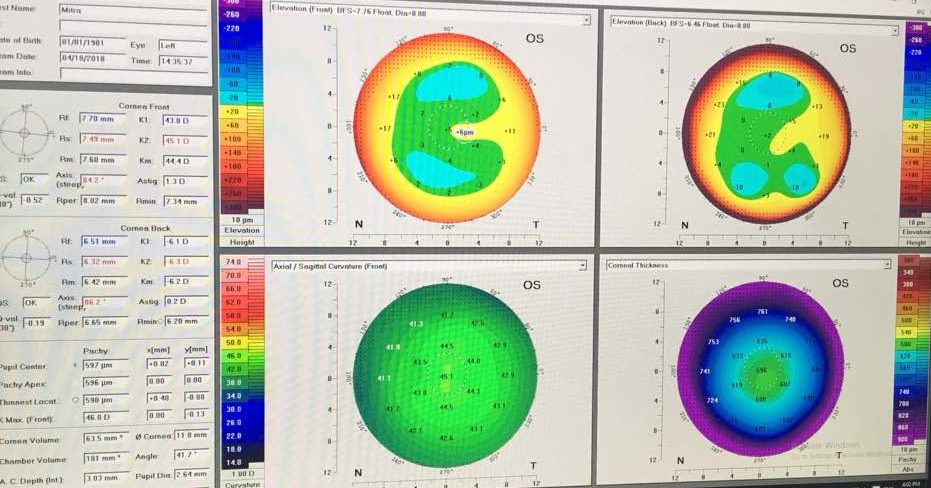
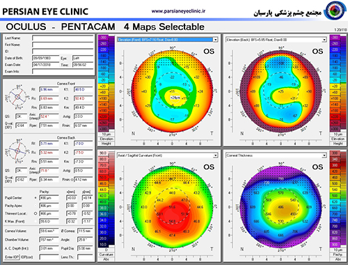
After measuring the peaks and troughs of the cornea surface through precise mathematical and optical calculations, the topography device imagines the corneal shape as a visual map. By looking at this map and its parameters, the physician will be able to screen those who are susceptible to have or already have keratoconus. In this map, the bulging points are represented by warm colors (red, yellow, …), while the notches are represented by cool colors (blue, etc.). In keratoconus, an unnatural bulging region in red is observed in the center of cornea.
In Parsian clinic, for preoperative examinations, several advanced devices are used:
Pentacam HR, Tomey, iTrace, Orbscam, Sirius and CASIA
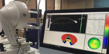
Pentacam topography and tomography device
Topographies have different types and are offered by various companies. One of these devices is Orbscan which has been under usage as the superior diagnostic technology for many years. In Parsian clinic, we have also used it over the past few years. However, recently a more advanced technology called Pentacam has been introduced. This device is very modern and advanced which in addition to the anterior surface of cornea, it also examines its posterior surface and depth. The cameras devised in this device use a very cutting-edge technology and operate very accurately. With this device, in addition to topography, one can also measure the corneal thickness at various points. Further, the depth of anterior room is also measured at different regions. Pentacam benefits from an advanced imaging technique called Sheimpflug, where the images captured by this system are more accurate and clearer than previous systems. This device has undergone various transformations and advances over years in terms of both software and hardware, and we always use the latest version of this device.
Concurrent with advanced centers of the world, Parsian clinic equipped itself with Pentacam, and we are the first center across the province and among the first countries that use Pentacam for performing topography. Usage of this device enhances the confidence level for performing LASIK operation.
In the images, we see a view of this device and one sample of the four maps of corneal topography.