
Angiography refers to imaging the blood vessels.
In this type of imaging, typically a harmless chemical known as contrast agent is injected through the veins to the patient, after which the image is captured. The contrast agent has the property to unveil the details of the blood vessels.
In ophthalmology, angiography is used for investigating the blood vistas of the retina and choroid, and the contrast agent for this purpose is called Fluorescein, and as such this angiography has been called after this agent. Retina and choroid have numerous blood vessels. In both ocular and non-ocular diseases, these vessels may be damaged, requiring this type of imaging for diagnosis and determining the extent of damage. One of the common and famous diseases affecting the blood vessels of the body including the blood vessels of the eyes is diabetes.
In diabetes, the blood vessels of the retina become blocked. Furthermore, the wall of the vessels also become fragile, causing bleeding and inflation inside the eye. If this condition is not diagnosed timely and treated accordingly, it results in diminished vision and even blindness.
Many people with diabetes may be asymptomatic for many years and their disease may be diagnosed by chance and by examining the deepest part of the eye. The deep eye examination as well as Fluorescein angiography cause diagnosis and determination of the extent of involvement of the eye. Based on that, the physician can determine the type and course of treatment.
In order to treat the vascular complications of diabetes in ophthalmology, laser is used. In order to get information about laser therapy, you can refer to the page Therapeutic Services in our website.
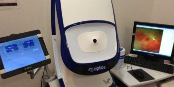
Today, angiography devices of the eyes have advanced considerably and digital techniques are used for imaging. We in Parsian clinic use the most advanced angiography device. This device which is called Optos is unrivaled in this regard and only a limited number of these device has been installed in the country. In the province, Parsian clinic is the only center equipped with this device.
The sharpness of imaging and field of measurement by this device are superior than measurements by other devices. It can image the retina and angiography with the angle of 210°. This value in devices reaches at most 50°.
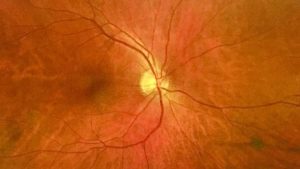
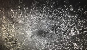
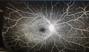
In the images, you can see a sample of angiography of retinal vessels in a normal individual and diabetic individual as well as open angle retinal colored imaging using this device.
Parsian subspecialty ophthalmology clinic is proud to say that it imported the technology of digital angiography of the eyes in 2006 for the first time, and since then it has always updated its technology.