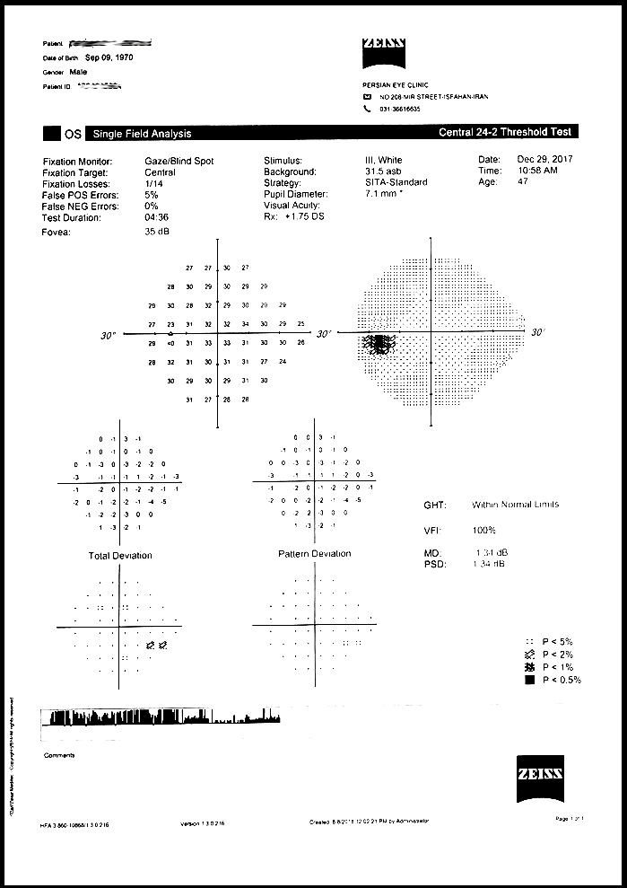
Perimetry means measuring the field of vision
Human vision is categorized into two parts: central vision and peripheral vision.
The central vision occurs when we directly look to something and recognize it. However, when we look at a point, we inevitably albeit unclearly see the points around it and sense it, as with when an object or person approaches us laterally. The peripheral vision is used for mobility and understanding the environment. The peripheral vision or field of vision of humans has dimensions and limits which can only be measured by special devices. The central vision determines the clarity of our vision as well as the quality of seeing colors, whose sensor is located in the retina Center. Unlike peripheral vision, measurement of the central vision is easy and can be measured via vision boards which are installed in all ophthalmology clinics. The central vision for near sight can also be measured by a vision board specifically designed for the near sight. In some disorders such as presbyopia, the near sight is impaired, while the far sight is absolutely normal. In myopia which is one of the common causes of vision weakness, the opposite occurs.
Some diseases first impair our peripheral vision and over time they also affect our central vision. The most important and common diseases include glaucoma. In this disease, first the peripheral vision is damaged and therefore up to the advanced stages of the disease the patient may not notice it, and when they notice it the peripheral vision has become very narrow and the patient may have found a tunnel like vision.
Via perimetry, the magnitude of the peripheral vision of the person is determined. This test would be very helpful for early diagnosis of glaucoma. Further, in a patient which is undergoing treatment, this test helps to understand whether the disease has been controlled or is progressing. In other diseases which involve the optic nerves or even brain nerves such as cerebral hemorrhage and tumors, this test helps in diagnosing the lesion.
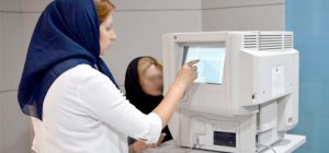
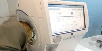
Concerning the procedure of this test, the patient should perceive a small illuminating object radiated on different parts of his or her retina in a semi-dark hemisphere. With changes in the lighting as well as the size of this object, the sensitivity of the retina environment and the extent of damage are measured.
This test alongside OCT of retina and optic nerve significantly contribute to diagnosing and treating glaucoma.
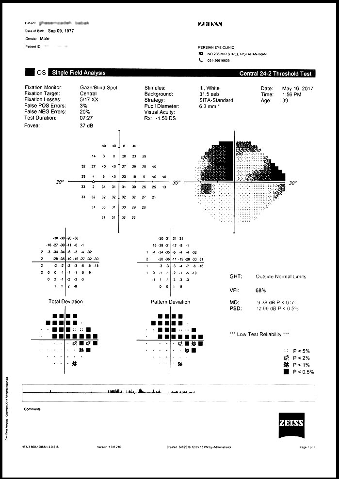
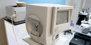
Persian subspecialty ophthalmology clinic benefits from these two advanced technologies to offer to patients.