
Other diagnostic services
in Persian Eye Clinic In addition to the diagnostic services you observed in other parts of this site, in this clinic, other ophthalmology diagnostic services are also presented which are summarized as follows.
Pachymetry
achymetry means measuring the thickness of cornea. This measurement is performed at different points and with technician and different devices numerically or as a colored map, where each color represents a special numerical value, such that with a quick glance to the map, we can guess the corneal thickness at every region or point.
Determination of corneal thickness is essential for decision-making on Lasik operations, because if the cornea is a thin, some limitations occur in the operation. In addition, in patients with glaucoma, today pachymetry is performed routinely, because it has been found that the corneal thickness is effective in determining the level of ocular pressure, where the thinner the cornea, the ocular pressure will be underestimated and the probability of error in measuring the ocular pressure and damage to division grows and vice versa; higher corneal thickness has a protective role in glaucoma patients. The pressure taken by the software of the device or calculation of the physician is adjusted based on the thickness of cornea whereby the actual ocular pressure is obtained.
Tonometry
Tonometry means measuring the intraocular pressure which is performed with different devices and methods. The most common method for measuring the ocular pressure is use of Applanation tonometer, which has been installed on ophthalmology examination devices.
One of the novel methods of tonometry is use of biomechanics device. Using this device, the role of confounding factors affecting the measurement is removed and the device measures the thickness of cornea concurrently, presenting the adjusted and actual pressure of the eyes. The most advanced available biomechanics device is called Corvis, which currently is only available in Parsian clinic and in a few number of other centers. For more information on biomechanics, click on it.
Specular microscopy
This method is used to investigate the quantity and quality of the internal layer cells of cornea known as endothelial cells. The human cornea has three major layers: the external layer (epithelium) has a protective and coating role causing uniform distribution of tears, whose thickness is around 50 miron. The middle layer known as the stroma constituents 90% of the corneal thickness, which gives the cornea its thickness, and finally the internal layer is called endothelium.
Endothelium is the most sensitive layer of cornea which is composed of one cellular layer, whose thickness is around 10 micron. The role of these cells is keeping the cornea transparent. These cells function as a two-sided pump adjusting the extent of fluid inside the stroma of cornea. Degradation of this cellular layer causes the cornea to inflate and become turbid necessitating corneal transplantation. The cause of development and degradation of endothelial cells is impact, surgical operations, and some genetic diseases. These cells cannot be viewed by unaided eye and even be typical devices, and can only be observed via special microscopy devices.
Parsian clinic is one of the first centers in the country to have provided this technology to citizens.
Measuring contrast sensitivity
One of the dimensions of human reason is perceiving contrast sensitivity. In addition to having good peripheral and central vision, we should also have a good contrast sensitivity, in order to be able to identify objects clearly and well. If we want to investigate contrast in colors, we should compare white with black. The contrast difference of these colors is maximum. If we place a black object on another piece with the same color, we cannot discern them, because the difference of their contrast is zero. However, if we place a white piece on the black piece, they can be easily differentiated from each other. The contrast of gray color is between black-and-white. Contrast sensitivity helps humans during the night and when the light is dim, and reduction of contrast results in diminished night vision. Different factors cause diminished contrast in the vision, including turbidity and lesions in visual tracts (especially cataracts) along with the complications of operations performed on the cornea. Excessive opening of the eye pupils in the evening and night causes diminished contrast and impaired recognition.
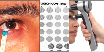
Four measurement of contrast, special tables are used, in which the color of texts and objects gradually fades. In addition, the size and distance of objects also decrease. At the minimum color and size the person can differentiate between two objects, the limit and extent of perception of contrast is obtained.
The figure demonstrates a sample of the device and the diagram of contrast curve used based on the device in Parsian clinic which is among the standard devices in this regard.
Investigating tears and lacrimal ducts
Tear is one of the important and vital functions of the eye. The tear is secreted from the lacrimal gland and once it scattered on the corneal and conjunctival surface, it leaves the lacrimal ducts and enters the throat, and this flows constantly. The main component of tear is water. There is also a thin lipid layer across the tear preventing its evaporation. In the deep part of tear, there is a layer of mucus causing adhesion of the aqueous layer of the tear to the corneal surface and homogeneous dispersion of the tear on the corneal surface. In addition to its moisturizing properties, tear has also several other materials thereby feeding and protecting the eyes against microbes. Therefore, deficient tear causes dryness and degradation of the surface of the eyes and increases the risk of infection and corneal ulcer.
Eye dryness has two major mechanisms: diminished secretion or increased outflow of tears from the eye. Diminished secretion occurs due to degradation of the lacrimal glands’ secretion, which is developed by different causes. Increased outflow occurs due to anatomical disorders of the eyelids and hyper-evaporation of tears. Today, environmental and occupational factors and overuse of computer and similar devices are among the causes of eye dryness.
To diagnose eye dryness, simple devices, examination with device, and sometimes complex devices are used. One of the ways is usage of spatial paper bands, where based on the extent of soaking the paper, the level of tears is measured. This test is called Shirmer test. Another way for diagnosing eye dryness is staining the surface of the eye with Fluorescein or other materials.
Another disorder of the lacrimal system is watery eyes. This condition is in contrast to eye dryness. The reason of the watery eyes is either hyper-secretion of tears or reduction of its outflow. Increased lacrimal secretions occur in response to irritating factors such as foreign body, impact, infections, and sensitivity. Another reason is blockage of outlet lacrimal ducts, which develops due to different reasons and the physician can diagnose the causes through examinations.
In Parsian clinic, the specialist and subspecialist ophthalmologists in particular in lacrimal system and duct are able to diagnose and treat different disorders of tears and lacrimal ducts.