Sonography is a type of imaging in which
ultrasound waves are used for development of the image. These waves collide with the tissue or organ of interest and the device analyzes the waves reflected from the organ of interest thereby specifying the normality or abnormality, and diagnosing the type of damage and visualizing it. The principles of sonography are similar to those of OCT (refer to the page of OCT), with the difference that since OCT has optical waves properties, it cannot cross turbid environments, and it is used only when the environment is transparent. However, soundwaves can also cross turbid tissues and environments, and unlike CT scan in which x-ray is used and is harmful to the human body, sonography has no risks and can even be used during pregnancy.
The resolution and diagnostic power of sonography depend on that wavelength and frequency of sound waves used in every device. The longer the sound wave (lower frequency), the greater the power of penetration of waves will be, thereby examining deeper tissues. However, the obtained images will have lower resolution, and in contrast the higher the frequency, the lower the penetrative power will be, but the diagnostic power and accuracy increases. Therefore, in ophthalmology if we want to investigate deeper layers of the eyes including retina and eye orbit, low-frequency sonography is used. On the other hand, if we want to examine the interior layers of the eye including cornea, the angle of the anterior chamber, lens, and the anterior parts of vitreous body, we use a higher frequency sound probe (lower wavelength). Furthermore, in sonography, different imaging techniques including A-Scan, B-scan or a mixture are used.
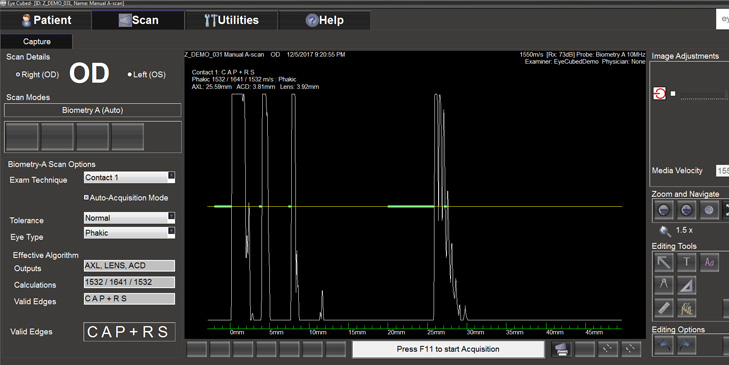
In the past, A-Scan sonography was used for performing biometry in all patients. However, today optical methods have replaced sonography. Nevertheless, still if the environment of the eye is not clear, such as when the cataract is concentrated or there is bleeding in the vitreous body, A-Scan sonography is used for parametric (Refer to perimetry page in our site).
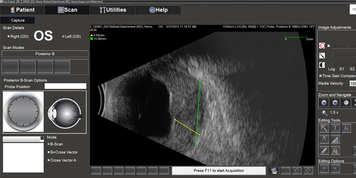
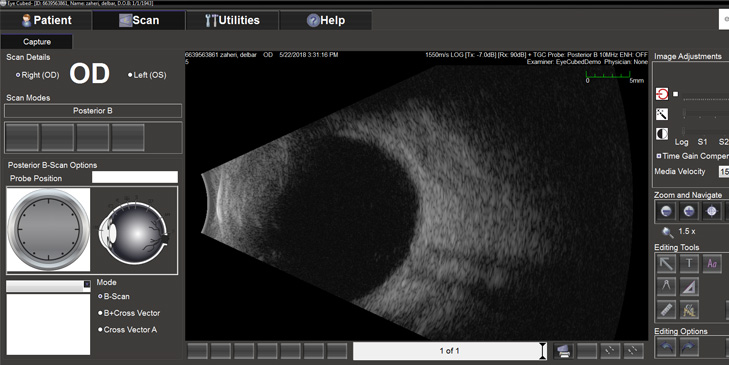
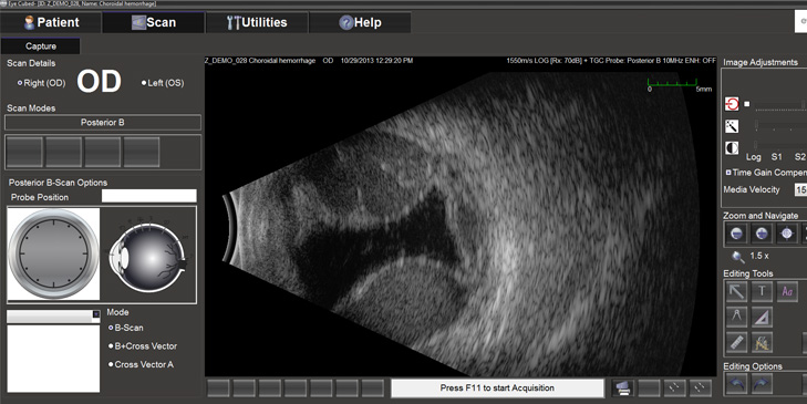
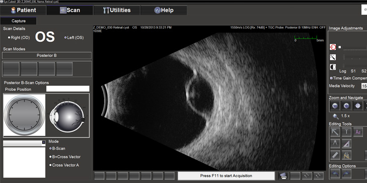
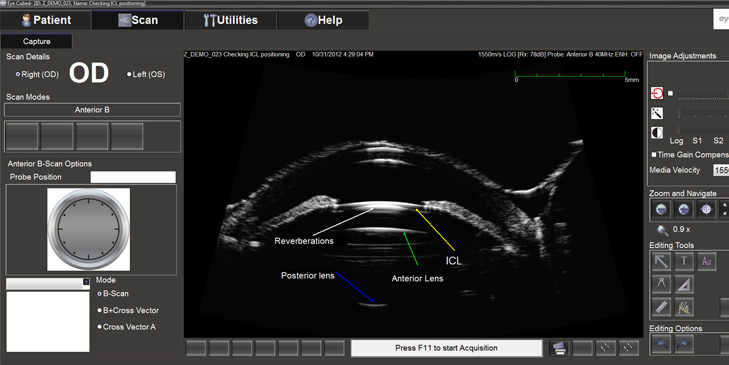
In B-scan, we observe a slice of the anatomical view of the eyes, and in A-scan we see the height of the wave reflected from each tissue as horizontal and vertical lines in two dimensions. The peak height of every wave represents each tissue of the eye. Since in sonography of anterior parts of the eye, high-frequency device and probe are used, a very clear anatomical image of this part is obtained which is known as ultrasound biomicroscopy (UBM).
In Parsian clinic, advanced and well-equipped devices of sonography with high-resolution and penetration are used to diagnose ocular lesions and for performing biometry. All of the three types of imaging including anterior, UBM, posterior (B-Scan), and biometry (A-scan) are performed based on the requirement of each patient.